Endometrial Metastasis from Breast Carcinoma Simulating a Primary Uterine Malignancy
Elias Hajal, Emile Dabaj, Mohamad Kassem, Elie Snaifer and Fatima Ghandour
Emile Dabaj1*, Elias Hajal1, Mohamad Kassem1, Elie Snaifer1 and Fatima Ghandour2
1Department of Pathology, Saint George Hospital University Medical Center, University of Balamand, Lebanon
2Department of Obstetrics and Gynecology, Saint George Hospital University Medical Center, University of Balamand, Lebanon
- *Corresponding Author:
- Emile D
Department of Pathology
Saint George Hospital University Medical Center
University of Balamand, Lebanon
Tel: +92-32483371653
E-mail: emile.dabaj@gmail.com
Received Date: May 17, 2017; Accepted Date: May 27, 2017; Published Date: June 03, 2017
Citation: Dabaj E, Hajal E, Kassem M, et al. Endometrial Metastasis from a Breast Carcinoma Simulating a Primary Uterine Malignancy. J Adenocarcinoma. 2017, 2:1. DOI: 10.21767/2572-309X.100016
Introduction
Metastases in the female genital tract occur from cancers that arise in various anatomic sites [1]. The ovary is the most common metastatic site followed by the vagina, cervix, uterine corpus, and fallopian tubes. Among extra genital cancers metastasizing to the uterine corpus, breast is the most common primary site [2]. Lobular carcinoma is the most common type of breast carcinoma that metastasizes to the uterus [3]. We herein report a case of uterine metastasis in a postmenopausal woman who had infiltrating lobular carcinoma of the breast.
Case presentation
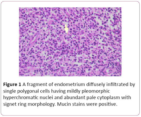
A 65-year-old woman was diagnosed 2 years ago with breast carcinoma. Right modified radical mastectomy and axillary lymph node dissection were performed. Pathological studies showed infiltrating lobular carcinoma signet type grade II (Bloom and Richardson), measuring 6-8 cm in its largest diameter, stage T3N2M0. All 22 axillary lymph nodes were involved with tumour. Immunohistochemistry revealed tumour cells to be negative for progesterone receptors (PR) and positive for estrogen receptors (ER) and CerbB2 positive by flow cytometry. Therefore, the patient received four cycles of adjuvant chemotherapy. Radiotherapy of 5000 cGy was also performed to the chest wall, right supraclavicular and right axillary areas and she was started on z which had cystic changes compatible with Tamoxifen-induced modifications. Hysteroscopy showed a hyperplastic endometrium. Endometrial curettage was performed to confirm the diagnosis of metastatic uterine carcinoma compatible with breast origin. The pathology report described a fragment of endometrium diffusely infiltrated by single polygonal cells (Figure 1) Similarly, Review of the previous breast tumour showed identical morphology confirming the diagnosis. A complete workup, including a normal level of CA 15-3 tumour marker, showed bone metastasis. The patient was treated with 10 cycles of Aredia (pamidronate).
Discussion
Breast cancer is the most common female cancer, the second most common cause of cancer related death in women, and the leading cause of death in women ages 40 to 59 [4]. Most common sites of metastasis of breast cancer are the lungs, followed by bones, liver, and brain whereas metastases in the female genital tract neraly are uncommon [2,3].
Metastatic disease to the female genital tract from the breast is rare. When it does occur, it mostly involves the ovaries possibly due to the nature of the stromal metabolic environment in terms of pH and oxygen tension. Loss of expression of the adhesive molecule E-Cadherin in infiltration lobular but not ductal carcinomas has been proposed to partially explain the differences in metastatic patterns [5,6]. Stemmer Mann proposed that majority of uterine metastases are a result of local lymphatic spread from earlier ovarian metastases as compared to probable hematogenous spread when the ovaries are disease-free [1].
Metastases to the uterus from extragenital locations account for less than 10% of all cases of metastases to the female genital tract (nearly 200 cases reported, more than half from a primary breast cancer) [7]. Within the uterus, the myometrial involvement predominates over endometrial (with stromal infiltration and sparing of the glands) and thus patients can be asymptomatic [8]. 43 autopsy cases and 9 hysterectomy specimens, and reported metastases in 96% of cases found in the myometrium with about a third also demonstrating endometrial metastases. Only 3.8% were limited to the endometrium [9]. Breast and colon cancers are the most frequent primary neoplasms found to metastasize to the uterus [10].
Metastases to the cervix are even rarer with 3 cases of isolated cervical metastasis being reported. The latest of which was documented by Andrew et al. [10]. A case of a 43 years old female who had a history of intraductal carcinoma of her left breast that was surgically managed 2 months prior to new onset abnormal vaginal bleeding. An endometrial biopsy performed as part of the workup revealed a poorly differentiated adenocarcinoma with immunohistochemical staining patterns similar to her previously diagnosed breast cancer. The patient was managed with a hysterectomy procedure which grossly revealed a fleshy polypoid endocervical mass. Microscopically, cords of carcinoma cells were identified, a pattern characteristic of breast carcinoma [11].
Eight cases of isolated uterine metastasis after breast cancer where infiltration rates of endometrium and myometrium were found to be comparable. The histologic type predominantly in their series was invasive lobular carcinoma [12]. For the past 2 decades, Tamoxifen has been a wellestablished drug in the management of both early and metastatic breast cancer. Risks of uterine cancer after its use, however, also have become well established, especially increasing after its long-term use [13].
Breast cancer metastases, particularly lobular, expresses cytokeratin 7 on immunohistochemistry most when compared to other extragenital metastasis. GATA-3 expression also reported as high in these cases. In our case report, the high clinical suspicion and the morphological resemblance to the slides obtained earlier from the breast carcinoma precluded the need to perform these tests.
In conclusion, it is prudent to entertain the possibility of uterine metastasis, whether isolated or in association with other sites in the female genital tract, from a breast cancer primary in patients presenting with vaginal bleeding after their breast cancer management. Tamoxifen use in itself predisposes to an array of conditions that can cause vaginal bleeding but its use doesn’t rule out a possible concomitant uterine metastasis from the mammary primary location. Metastatic carcinoma to the endometrium may be suspected if one of the following signs is present:
1. A tumour with an unusual gross or histological pattern for primary endometrial carcinoma.
2. Diffuse replacement of endometrial stroma with preservation of endometrial glands.
3. Lack of accompanying premalignant changes in the residual benign endometrium.
4. Lack of tumour necrosis.
Acknowledgments
Snaifer Elie, MD, Assistant Professor of Obstetrics and Gynecology, Faculty of Medicine, University of Balamand. Ghandour-Hajj Fatima, MD, Assistant Professor of Pathology, Faculty of Medicine, University of Balamand, Lebanon.
References
- Stemmermann GN(1961)Extrapelvic carcinoma metastatic to the uterus. Am J ObstetGynecol 82: 1-1266.
- Kumar NB, Hart WR (1982)Metastases to the uterine corpus from extragenital cancers. A clini study of 63 cases.50: 2163-2169.
- Mazur MT, Hsueh S,Gersell DJ (1984)Metastases to the female genital tract. Analysis of 325 cases, Canc 53: 1978-1984.
- Jemal A, Siegel R, Ward E, Hao Y, Xu J, et al. (2009) Cancer statistics. CA Cancer J Clin 59: 25-249.
- Sastre-Garau X, Jouve M, Asselain B (1996) Infiltrating lobular carcinoma of the breast. Canc77: 113-20.
- Moll R, Mitze M, Frixen U, Birchmeier W(1993) Differential loss of E cadherin expression in infiltrating ductal and lobular breasts carcinomas. Am J Pathol 143:1731-42.
- Silverberg SG, Kurman RJ (1992) Atlas of Tumor Pathology: Tumors of the Uterine Corpus and Gestational Trophoblastic Disease. Armed Forces Institute of Pathology, Washington.
- Maymon R, Czernobilsky B, Lifschitz-Mercer B, Schneider D, Bukovsky I, et al.(1996) Metastatic breast carcinoma manifesting as postmenopausal uterine bleeding in a patient on tamoxifen therapy. Eur J GynecolOncol17: 319-21.
- Neelam B, William R,(1982) Metastases to the Uterine Corpus from Extragenital Cancers A Clinicopathologic Study of 63 Cases.Canc 50: 2163-2169.
- Andrew EG, Charles B, Chad M, Jerome B (2004) Isolated cervical metastasis of breast cancer: a case report and review of the literature. GynecolOncol95:267-269.
- Le Bouëdec G, Kauffmann P, De Latour M, Fondrinier E, Curé H,et al.(1991) Centre Jean Perrin, Clermont-Ferrand. Jof GynecolObste and Biol of Reprod 20:349354.
- Bernstein L, Deapen D, Cerhan JR,(1999) Tamoxifen therapy for breast cancer and endometrial cancer risk. J Natl Cancer Inst 91: 1654-62.
- Euscher E, Malpica A (2014) Use of immunohistochemistry in the diagnosis of miscellaneous and metastatic tumors of the uterine corpus and cervix. SeminDiagnPathol 31 :233-57 4.

Open Access Journals
- Aquaculture & Veterinary Science
- Chemistry & Chemical Sciences
- Clinical Sciences
- Engineering
- General Science
- Genetics & Molecular Biology
- Health Care & Nursing
- Immunology & Microbiology
- Materials Science
- Mathematics & Physics
- Medical Sciences
- Neurology & Psychiatry
- Oncology & Cancer Science
- Pharmaceutical Sciences

